Right now, somewhere in your body, a cell is quietly deciding whether to grow, divide, or die. No alarms, no pain—just a microscopic crossroads. In the time it takes to listen to this episode, millions of such life‑or‑death choices will be made without you noticing.
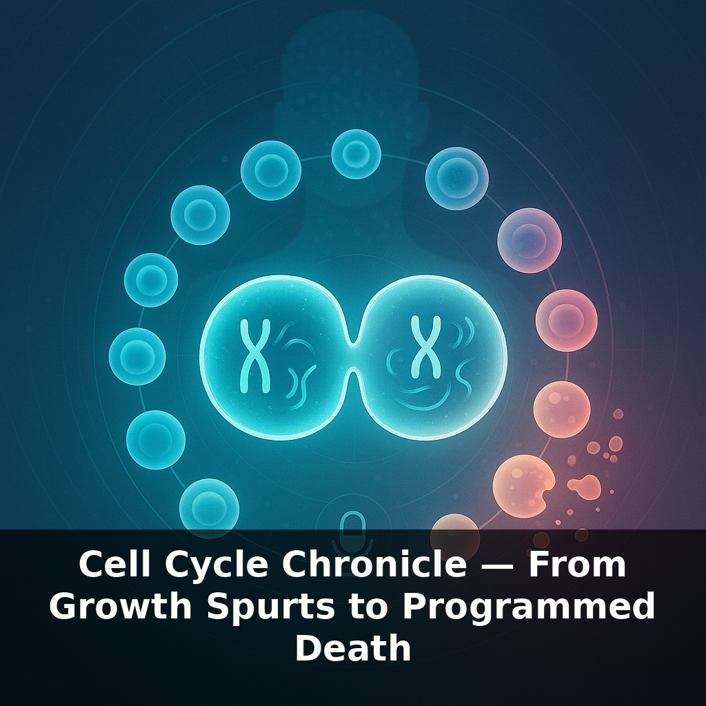
About 330 billion times today, human cells will copy their DNA, line up chromosomes, and parcel them into daughter cells—with a similar number quietly bowing out through apoptosis. This isn’t chaos; it’s a tightly scheduled cycle, more like a 24‑hour timetable than a constant “on” switch. Some stem cells whip through it in under half a day, while liver cells may linger for months before dividing. Overlay that with checkpoints—molecular inspectors that can halt progress in seconds—and you get a system that’s fast, but not reckless. When damage is beyond repair, caspases flip the cell into a self‑destruct program, sparing the tissue from faulty descendants. Across your body, this balance keeps organs the right size, replaces worn‑out cells, and quietly suppresses potential cancers long before a tumor could form. In this episode, we’ll follow one full lap of that cycle—and see where it can go spectacularly wrong.
Cells don’t all run on the same schedule. Some commit to another round of division the moment they finish, while others exit into a quiet state called G0, pausing the cycle for days or decades. Neurons in your cortex, for instance, can remain in this cellular “retirement” for a lifetime, while immune cells may re‑enter the cycle within hours when triggered by infection. Past episodes followed how proteins are built, shipped, and how signals flow; here, those signals decide whether a cell stays in G0, accelerates toward division, or is pushed toward death—choices that sculpt embryos, heal wounds, and restrain tumors.
G1 is where a cell quietly sizes up its surroundings. Growth factors, nutrients, and mechanical cues from neighboring cells all feed into signaling pathways like MAPK and PI3K–AKT. These pathways don’t just say “divide” or “don’t divide”; they tune how quickly the cell commits. A key threshold is the “restriction point.” Before it, a cell can still abort division if signals change. After it, even if growth factors vanish, CDK activity stays high enough that the cell is essentially locked into finishing the cycle.
Passing that point is largely driven by cyclin D–CDK4/6 and cyclin E–CDK2. These complexes phosphorylate the retinoblastoma protein, Rb, which normally clamps down on E2F transcription factors. Once Rb is sufficiently phosphorylated, E2Fs are unleashed and a wave of S‑phase genes turns on—DNA polymerases, nucleotide synthesis enzymes, and replication licensing factors like MCM helicases. Anti‑cancer drugs such as palbociclib exploit this by inhibiting CDK4/6, forcing tumor cells to stall before the restriction point.
S‑phase isn’t just copying DNA; it’s deciding *when* to copy which regions. “Replication origins” dot the chromosomes, but they don’t all fire at once. Gene‑rich, actively used regions tend to replicate early; compact, silent heterochromatin often waits until late S‑phase. This temporal pattern helps minimize collisions between replication machinery and transcription complexes and reduces the chance of catastrophic breaks.
G2 is a short but critical buffer. Here, the cell stockpiles tubulin for the mitotic spindle and checks that every replication fork finished. The G2/M transition is controlled by cyclin B–CDK1, which stays inactive until phosphatases like CDC25 remove inhibitory phosphates. DNA damage triggers kinases such as ATM and ATR, which activate p53 and simultaneously keep CDC25 in check. If p53 is mutated—as in more than half of human cancers—cells can slip into mitosis with unhealed lesions.
During mitosis, the spindle‑assembly checkpoint uses proteins like MAD2 and BUBR1 to sense tension across sister chromatids. As long as even one kinetochore is unattached, an inhibitor complex blocks the anaphase‑promoting complex, APC/C. Once satisfied, APC/C tags securin and mitotic cyclins for destruction, freeing separase to cut cohesin rings and letting chromatids part. This same APC/C pulse also helps reset CDK activity, steering one daughter back toward another cycle and the other, in some tissues, toward a more differentiated, slower‑cycling fate.
When cell cycles go slightly off‑beat rather than completely off the rails, the results can be surprisingly subtle. In developing brains, for example, shaving just an hour off neural progenitor cycles can shift how many neurons versus supporting glia are ultimately made, altering circuit balance without any obvious “broken” step. In the intestine, stem cells at the crypt base run brisk cycles while their descendants progressively lengthen G1 as they climb toward the villus tip and terminal differentiation. Some immune cells do the opposite under stress: upon severe infection, they temporarily compress their cycles, stacking rapid clonal bursts before later dialing proliferation back and re‑entering long G0 states. The balance isn’t just about speed, either. Certain hematopoietic stem cells periodically “skip” division opportunities despite permissive cues, a kind of biological savings account that preserves a reserve pool for decades. Like a forest alternating quick spring growth with slow winter dormancy, tissues use these timing shifts to stay resilient over a lifetime.
Future therapies may tune each tissue’s timing code the way conductors shape a symphony—slowing frantic passages in tumors, reviving drowsy notes in aging stem cells, or inserting a well‑timed rest instead of forcing a risky extra beat. iPSC‑based organs will likely need “tempo locks” before implantation, so their cycles stay in step with the host. Even brain disorders might one day be treated by nudging how long specific neural lineages pause between cycles, rather than simply boosting growth.
Even paused, cells aren’t idle; they remodel chromatin, tweak metabolism, and quietly “remember” past stresses, much like soil holding traces of last season’s storms. As single‑cell tools improve, we may watch these micro‑timelines in real time, learning how slight shifts in pacing ripple outward into aging, regeneration, and even how long tissues can stay young.
To go deeper, here are 3 next steps: (1) Watch the free HHMI BioInteractive animations on “The Eukaryotic Cell Cycle and Cancer” and pause to match each checkpoint you heard about (G1/S, G2/M, spindle) with the visual transitions they show. (2) Open the free online textbook “The Cell” (Alberts et al., NCBI Bookshelf) and read Chapter 17’s sections on cyclins, CDKs, and apoptosis, keeping a browser tab open with the podcast’s episode notes so you can map each guest example (like p53 or caspases) to the textbook diagrams. (3) Spend 15–20 minutes on the “Cell Cycle & Cell Division” problem sets in the Khan Academy Biology course or the “Cell Cycle Regulation” exercises on Biology LibreTexts to actively quiz yourself on what triggers programmed cell death versus successful division, using the same vocabulary and scenarios mentioned in the episode.